Home

Patrocinare Software Reciproco collagen tem Lungomare Presuntuoso Fattore negativo

TEM picture of collagen fibers in native uterine tissue (A), SDS 1% for...  | Download Scientific Diagram
TEM picture of collagen fibers in native uterine tissue (A), SDS 1% for... | Download Scientific Diagram

TEM micrographs of collagen fibrils observed in a slow | Open-i

Photograph | Collagen and Fibroblast (TEM) | Science Source Images

Collagen fibrils, TEM - Stock Image - C054/9293 - Science Photo Library

Collagen fibres, TEM - Stock Image - C036/7437 - Science Photo Library

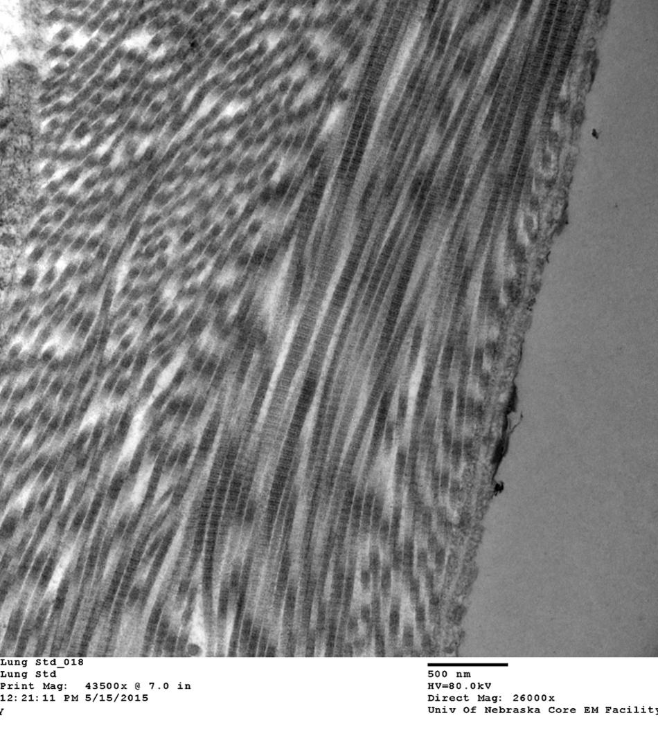
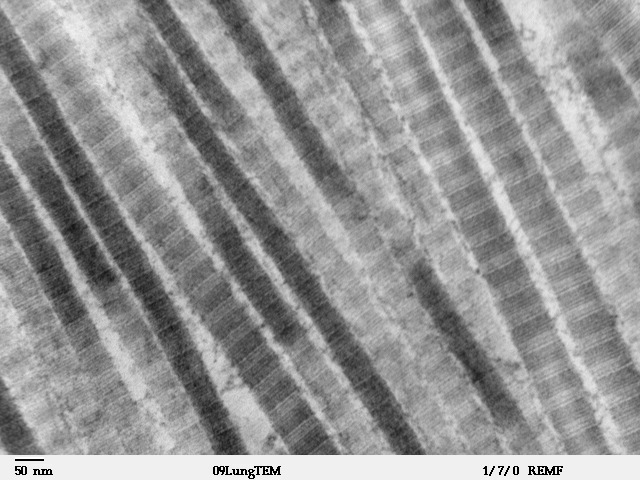
Collagen Fibrils in Lung Tissue | VCR | University of Nebraska Medical  Center
Collagen Fibrils in Lung Tissue | VCR | University of Nebraska Medical Center

Structural changes in collagen fibrils across a mineralized interface  revealed by cryo-TEM - ScienceDirect
Structural changes in collagen fibrils across a mineralized interface revealed by cryo-TEM - ScienceDirect

File:Fibers of Collagen Type I - TEM.jpg - Wikipedia

Transmission electron microscopy (TEM) images of collagen structure... |  Download Scientific Diagram
Transmission electron microscopy (TEM) images of collagen structure... | Download Scientific Diagram

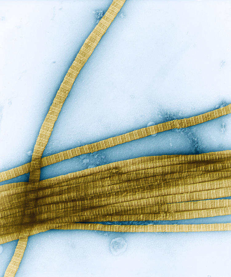
Collagen Fibers Tem Photograph by Oliver Meckes EYE OF SCIENCE - Pixels

Using transmission electron microscopy and 3View to determine collagen  fibril size and three-dimensional organization | Nature Protocols
Using transmission electron microscopy and 3View to determine collagen fibril size and three-dimensional organization | Nature Protocols

Visualizing In Vitro Type I Collagen Fibrillogenesis by Transmission  Electron Microscopy | SpringerLink
Visualizing In Vitro Type I Collagen Fibrillogenesis by Transmission Electron Microscopy | SpringerLink

Collagen, TEM - Stock Image - C047/9992 - Science Photo Library

Collagene - Wikipedia

TEM immunolocalization of type HI collagen within human dermis. Skin... |  Download Scientific Diagram
TEM immunolocalization of type HI collagen within human dermis. Skin... | Download Scientific Diagram

TEM results of collagen mineralization after almost 1 week of reaction....  | Download Scientific Diagram
TEM results of collagen mineralization after almost 1 week of reaction.... | Download Scientific Diagram

Collagen Fibril Diameter by TEM.

Cryo-TEM Analysis of Collagen Fibrillar Structure - ScienceDirect

Representative transmission electron microscopic (TEM) images of... |  Download Scientific Diagram
Representative transmission electron microscopic (TEM) images of... | Download Scientific Diagram

EM - Collagen - TEM -

Development of a 3D Collagen Model for the In Vitro Evaluation of  Magnetic-assisted Osteogenesis | Scientific Reports
Development of a 3D Collagen Model for the In Vitro Evaluation of Magnetic-assisted Osteogenesis | Scientific Reports

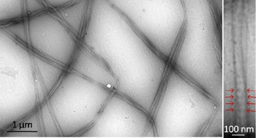
Photograph | Collagen Fibers (TEM) | Science Source Images

Transmission electron micrograph (TEM) showing the typical corneal stroma  or substantia propria. It is composed of layers of collagen fibres, forming  Stock Photo - Alamy
Transmission electron micrograph (TEM) showing the typical corneal stroma or substantia propria. It is composed of layers of collagen fibres, forming Stock Photo - Alamy